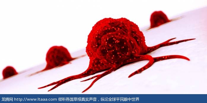
新型显微镜利用光线来“洞穿”组织样本以寻找癌变 [美国媒体]
乳房肿瘤切除手术的终极目标,是从人的胸部切除所有的癌变组织,并同时尽可能保留更多的健康组织。然而这事说起来容易做起来难。由于当前的科技水平还达不到在显微镜下观察组织的程度,因此目前还无法分辨肿瘤周围区域是否含有癌变细胞。
A new microscope uses light to 'cut' through tissue samples and find cancer
新型显微镜利用光线来“洞穿”组织样本以寻找癌变
The ultimate goal of a lumpectomy is to remove all the cancerous tissue from a person's breast while saving as much of the healthy tissue as possible.
乳房肿瘤切除手术的终极目标,是从人的胸部切除所有的癌变组织,并同时尽可能保留更多的健康组织。
But that's even harder than it sounds.
然而这事说起来容易做起来难。
Without the ability to look at tissue under a microscope, it's currently impossible to tell whether the area surrounding a tumor contains cancerous cells or not.
由于当前的科技水平还达不到在显微镜下观察组织的程度,因此目前还无法分辨肿瘤周围区域是否含有癌变细胞。
Surgeons deal with this by taking samples from around the removed tumor, waiting for a pathologist to look at them, and performing additional surgeries if they turn out to be cancerous.
为达此目的,外科医生通过对已切除的肿瘤周围区域进行采样,然后再由病理学医生进行观察。如果观察的结果是有癌变,则再次进行手术。
This is not only time consuming and expensive, but also extremely stressful for the patient.
这种方法很费时而且费用昂贵,此外还会给病人造成很大压力。
A new microscope developed by researchers at the University of Washington might change that.
现在,一种由华盛顿大学的学者们发明的新型显微镜诞生了,这可能会改变上述情况。
It uses a super powerful light to optically "cut" through the area and see what's inside in around 30 minutes, so that surgeons can know whether that tissue sample still contains cancerous cells.
该显微镜使用一种超强光线,在视觉上得以洞穿肿瘤周围的组织并可在30分钟内观察其内部情况,这样一来外科医生就能得知该组织样本是否仍含有癌细胞。
If it does, they can go back and remove that tissue, hopefully getting rid of the cancer in one go. The researchers published their work this week in the journal Nature Biomedical Engineering.
若发现癌细胞,外科医生就可以回头做手术切除该组织,这样就有希望一次性切除癌变组织。学者们本周在《自然-生物医学工程》杂志上发表了他们的研究成果。
Unlike other areas of medicine, pathology hasn't advanced all that much.
和其他医学领域不同,病理学还没有如此发达。
To see if a piece of tissue is cancerous, pathologists still need to go through extensive preparation processes—like staining, slicing, and forming tissues into waxy blocks before they can look at them under a microscope.
为了查看一片组织是否发生癌变,病理学家仍旧需要进行大量的准备过程,比如染色、切片、然后把组织放到蜡块中成形,最后才是在显微镜下观察。
That process can take days, and it also limits the number of diagnostic procedures you can carry out on a single sample.
该过程可能花费数天时间,并且在一个组织样本中能够使用的诊断方式其数量也是有限的。
Once something is sliced and stained, it can't be retested for another disease or sent for genetic testing, which is an important part of cancer treatment.
一旦某组织样本被切片和染色了,它就不能再被用来测试其他疾病或者用来做基因检测,而基因检测在癌症的治疗中又是非常重要的一部分。
Because the new microscope design works quickly and without wrecking tissue, the surgeons can get results during the surgery itself, determine what needs to be done next, and save samples for other tests.
现在,由于新型显微镜在医疗过程中能够快速起到作用并且不用再破坏组织,因此外科医生得以仅通过外科手术就知道结果,以决定下一步该做什么,并且样本还可以被保留用来做其他检测。
The researchers say they are currently testing out the microscope on breast cancer samples, but in the future they plan to use it on other cancers that also involve solid tumors.
研究者们声称,他们正在乳腺癌样本上测试该显微镜,并希望将来能够将其用于包括实体瘤在内的其他癌症上。
Even further into the future, they hope to incorporate machine learning algorithms to help pathologists identify cancer more quickly and accurately.
甚至更远的将来,他们希望吸纳机器学习算法来帮助病理学家更快和更精确地找到癌变。
版权声明
我们致力于传递世界各地老百姓最真实、最直接、最详尽的对中国的看法
【版权与免责声明】如发现内容存在版权问题,烦请提供相关信息发邮件,
我们将及时沟通与处理。本站内容除非来源注明五毛网,否则均为网友转载,涉及言论、版权与本站无关。
本文仅代表作者观点,不代表本站立场。
本文来自网络,如有侵权及时联系本网站。
图文文章RECOMMEND
热门文章HOT NEWS
-
1
Why do most people who have a positive view of China have been to ...
- 2
- 3
- 4
- 5
- 6
- 7
- 8
- 9
- 10
推荐文章HOT NEWS
-
1
Why do most people who have a positive view of China have been to ...
- 2
- 3
- 4
- 5
- 6
- 7
- 8
- 9
- 10


